


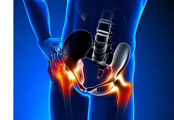
Hip joint osteoarthritis (coxarthrosis)- This is a chronic degenerative joint disease, which leads to the deformation of bone tissue.With cookardrosis, all joint components are involved in the pathological process: joint cartilage, bone structures adjacent to cartilage, synovial shell, ligaments, capsule and adjacent muscles.In the case of the disease, joint cartilage is destroyed, bone and osteophytes micro-re-re-laddimitations (bone growth) appear, and an inflammation of the muscle-ligamment of the hip joint is produced.
In the world, every fifth person complains about joint problems with the joints.This can be both pain and restriction of the joint movement and a combination of these symptoms.Every second outpatient vision falls to patients with bone muscle disorders, while 66 % of cases are people under 65.According to the latest epidemiological research, the prevalence of the osteoarthritis of the knee joints and hip among the adult population is 13 %.
Risk factors for coxarcher development:
- Genetic predisposition.A common cause of cookyrosis of the hip joints is the congenital or acquired mutation of the type of type II.
- Elderly age.The probable cause of the prevalence of osteoarthritis in old age is a discrepancy between the harmful effect on the articular cartilage of the external environment and its abilities to restore.
- Floor.Women suffer from osteoarthritis more frequently than men.This is due to the effects of the influence of female sexual hormones of estrogen in the minimum bone metabolism.However, the influence of the floor is ambiguous: according to some authors, unlike damage to other joints, there are no differences in the sexual base for cookcartosis: in men, the osteoarthritis of the hip joint is as often as in women.
- Excess body weight.The relationship is tested between excess body mass and the appearance of osteoarthritis.Excess adhesive tissue increases the harmful load in the cartilage.In addition, adipose tissue produces pro -inflammatory enzymes that damage cartilage tissue.
- Frequent development of bones and joints.According to the studies, 80 % of coxarchesis, which occurs without apparent reason, is associated with defects previously diagnosed in the development of the hip joint: dysplasia and subluxation.
- Heavy physical work.An excess load in hip joints with certain types of physical work can cause damage to the joints and the formation of osteoarthritis.At risk are agricultural workers, excavators and similar work specialties.
- InjuriesThe risk of developing coxarthrosis increases after an injury in the hip joint.In addition, both an injured and both articulation can be involved in the process.
- Professional practicing sports.Professional sport can cause the appearance of coxarthrosis both due to excessive load in the joints and injuries.Potentially dangerous sports include heavy athletics, athletics jump, parachute sport.
- Bones and joint diseases- Rheumatoid arthritis, psoriatic arthritis, joint infections, avascular necrosis, gouty arthritis, etc.
- Endocrine pathologies- Hypothyroidism, hypoparathyroidism, acromegaly (deteriorated function of the anterior pituitary gland), diabetes, obesity.
If similar symptoms are detected, consult a doctor.Do not self -medicate, it is dangerous for your health!
Symptoms of osteoarthritis of the hip joints
The main symptoms of coxarthrosis include: pain, mobility restrictions and crunch in the joints, their deformation, functional shortening of the lower limb and periodic swelling in the joints.
Pain of various intensity.Articulation pain is initially insignificant and arises for a short time.They appear or intensify while walking or with another physical effort, for example, during the squats, inclinations and weightlifting.As the disease develops, pain intensifies and even a long break does not bring relief.In addition, pain occurs with prolonged immobility and joint fixation in a position.
Patients complain about the "initiated" pain called in the hip joints after sleeping, driving in a car and another prolonged immobility.The "start" pain for coxarchesis does not last more than 30 minutes.The pain intensifies during hypothermia or in a stressful situation.They can be located in the area of the buttock or the groin, on the front or side surface of the thigh.With the spread of pain on the nerves of the lumbar plexus, it can be transmitted to the thighs away from the center of the body or in the knee.Sometimes, pain applies to lumbosacra column and coxis.
Joint mobility restriction.The movements in the hip joint with cookyrosis are limited due to pain.At the same time, rotation (tours both inside and outside) and that carry the lower limb (movement to the center of the body) are more frequently altered, but can be limited (movement from the middle axis of the body), as well as flexion and extension.The inability to make passive movements in the joint due to a pronounced pain syndrome causes compensatory pelvic bias.The patient's march changes, the buttocks stand out, the body deviates forward when transferring the weight to the damaged side.With bilateral damage in patients with cookardosis, a "duck march" is formed.
With coxarchesis it occurs periodicallySwelling in the jointwhich can be invisible due to the muscle layer and fat.In addition, the disease is characteristicCryst in the joints during the movement, its gradual deformation and functional shortening of the lower limb.
Often, an articulation is affected with the disease, so the process applies to others.But sometimes osteoarthritis affects several joints at the same time and is caused byotritis.Polyosteoarthrosis is characteristic of older people or a hereditary predisposition and concomitant diseases: bone diseases, joints and endocrine disorders.
Pathogenesis of the arthrosis of the hip joints
In the pathogenesis of the osteoarthritis of the hip joints, an important role for mechanical damage (injuries and microtraumas due to the increase in physical effort in the joint) and genetic factors, hormonal and metabolic, is played.It is often not possible to discover which factor has influenced the development of the disease in a particular patient, but often the disease develops after tissue damage with mechanical lesions.
The damage to the tissue stimulates the division of cartilage tissue cells (chondrocytes), while the production of proinflammatory cytokines increases, which are normally present in the cartilage in just small quantities.Citocins launch the inflammatory process, for example, under the influence of the proinflamatory cytocin IL-1, enzymes are distinguished that destroy the cartilage of the joint.In addition, under the influence of cytokines, the production of the Tsog-2 enzyme and other substances that have a toxic effect on the increase in cartilage.
The Synovites also play an important role in the development of coxarchery: inflammatory diseases of the synovial cover of the joints or ligaments with the accumulation of fluid in the cavity.
A decrease in elasticity and strength of articular cartilage associated with metabolic disorders leads to a decrease in their resistance to mechanical stress.With cookardosis, all joint components are involved in the pathological process, including a subchondral bone.Due to the fact that the large joints of the lower extremities explain the large joints of the body, experience significant mechanical stress, due to which microvalomas occur in the subcondral plate and cartilage.As a result of microvelomas, the subcondral bone is compact, which leads to the regional growth of bone tissue osteophytes.And this, in turn, stimulates a greater degradation of the articular cartilage.
In some cases, the osteoarthritis of the hip joint is inherited.Hereditary osteoarthritis is supposedly polygenic inheritance, due to the action of many genes, each of which affects weakly.The cause of some diseases is a mutation in genes that encode macromolecules of the joint cartilage, which causes its ruptures.The genes responsible for the division of the chondrocytes can also suffer.In addition, metabolic disorders are inherited, such as pyrophosphate arthropathy, a disease in which calcium pyrophosphate crystals accumulate in joint cartilage and synovial fluid.
Classification and stages of development of the articulations of the hip
Depending on the causes of the disease, coxarthrosis is divided into two main forms: primary (idiopathic) and secondary (which arises from or other diseases).
Primary Coksarrosis:
- Located (only affects hip joints):
- unilateral;
- bilateral.
- Generalized (pots.
Secondary osteoarthritis:
- Post -traumatic:
- Acute: as a consequence of acute injury;
- Chronic: due to classes of some sports or as a result of professional activity.
- Metabolic diseases (oponosis, hemochromatosis, Wilson's disease, Gaucher's disease).
- Congenital pathologies and development defects (congenital dysplasia of the hip joint, belongs to the disease, sliding of the femur epiphases, hypermobility syndrome, shortening of the lower limb, scoliosis, bone dysplasia).
- Endocrine pathologies (acromegaly, hypothyroidism, diabetes mellitus, hyperparathyroidism, obesity).
- Calcium rooms (pyrophosphate arthropathy, calcification tendonitis).
- Bone diseases and joints (rheumatoid arthritis, psoriatic arthritis, pedastic disease, avascular necrosis, infections).
According to clinical manifestations, the following forms of coxarcheis are distinguished:
- Little symptom.
- Manifesto, manifested by brilliant clinical symptoms:
- quickly progressive, in which symptoms develop in the first four years since the beginning of the disease;
- Slowly progressive: clinically significant symptoms appear after five years of the disease course.
According to image X -ray, two types of osteoarthritis of the hip joints can be identified:
- Hypertrophic: with signs of greater repair response (the lesions are replaced by a new tissue, for example, osteophytes appear);
- Atrophic (decrease in tissue volume).
The stages of the disease can be determined radiologically and clinically.To determine the radiological stage of the osteoarthritis of the hip joint, the classification of Kellgren and Lawrence (1957) is used more frequently.
Stages of osteoarthritis in the radiological classification
| Scenery | Signs |
|---|---|
| 0 | There are no signs of osteoarthritis in X -ray images |
| 1 | The articulation gap is not changed, individual regional osteophytes are displayed |
| 2 | The articular gap is not changed, significant regional osteophytes are displayed |
| 3 | The height of the joint space is moderately reduced, significant regional osteophytes are displayed |
| 4 | The height of the joint gap is significantly reduced, significant regional osteophytes and subchondral osteosclerosis (compaction of the bone tissue below the lower surface of the cartilage with the cartilage structure) are displayed) |
To determine the clinical stage of the disease, classification (1961) is used, which uses clinical signs and visualization criteria.
Clinical stages of osteoarthritis
| Scenery | Signs |
|---|---|
| 0 | The articular gap is unequivocally reduced and unequally, the edges of the joint cracks are slightly pointed (initial osteophytes), a slight restriction of the movements is observed |
| 1 | The articular gap is significantly reduced (50-60 %), significant osteophytes, subchondral osteocosclerosis and cystic lighting in bone epiphases;The clinic is predominated by the restriction of joint mobility, a rough crunch during movements, insignificant or moderate muscular atrophy |
| 2 | deformation, stiffness of the joint;The articular gap is reduced by more than 60-70 % of the standard or complete |
Complications of the arthrosis of the hip joints
With coxarthrosis, all complications are associated precisely with pathological changes in the joints.
The course of cookyrosis can be complicated by local inflammatory processes:
- Bursite - inflammation of synovial bags in the joints;
- Tendovaginitis: inflammation of the inner layer of the vagina of muscle tendons;
- Nervous tunnel syndrome pins due to the formation of large osteophytes or joint deformation.
With the progression of coxarchtosis and its transition to clinical stages II and III, pain limits the mobility of the articulation, and over time, joint ankylosis (fibrous, bone or cartilage) occurs, accompanied by its complete immobility.
Significant joint deformation can lead toFractures or aseptic bone necrosis.For cookyrosis, aseptic necrosis of the femoral head is the most formidable complication.
With pronounced cooksis, it can occursubluxation and dislocation of the jointas well as the penetration of the femoral head in the pelvic cavity.Dyslocations and subluxation of the hip joint lead to pain (at the acute, then opaque and sore principle), intensifying during the walk and other physical effort, as well as to the deformation of the joint, lame and, sometimes, to shorten the affected limb.
Despite the lack of systemic manifestations of osteoarthritis, in modern clinical practice, more attention is paid to the diseases associated with it.These are such pathological conditions that exist or arise in the context of current disease.In relation to the inflammatory reactions that arise during osteoarthritis, the formation of atherosclerotic plaques is improved on the internal walls of the vessels, which increases the riskCardiovascular diseases.A decrease in physical activity due to pain and restriction of joint mobility leads toObesity, depression and deterioration in the quality of life.With prolonged use of non -steroidal anti -inflammatory drugs,The upper gastrointestinal sections are affected,And alsoThe risk of cardiovascular pathologies and kidney diseases increases.
Diagnosis of articulations of the hip joints
The diagnosis of "cooksarrosis" is performed on the basis of clinical manifestations and radiological examination.There are no characteristic laboratory signs for the diagnosis of osteoarthritis.
Among the clinical manifestationsThe main one for the diagnosis of osteoarthritis of the hip joint is pain and its character.The pain for the osteoarthritis of the hip joint occurs and grows gradually for several years (sometimes several months with a rapidly progressive way).Pain occurs or improves during physical effort or standing position.If the patient begins to feel pain alone, then inflammation (synovitis) joined.The statement is observed up to 30 minutes in the morning and with prolonged immobility.
The limitation of joint mobility is gradually increasing, this applies to both active and liabilities.With the development of the disease, the joints are deform, the functional shortening of the length of the limb can occur.
In a physics examThere is a limitation of joint mobility, its deformation, shortening of the limbs, pain in the palpation of the joint and a great turn of the femur, muscle atrophy.
Laboratory methodsThe diagnosis of osteoarthritis of the hip joints is not required.However, they can be used for the differential diagnosis of coxarthrosis with arthritis (rheumatoid and chronic), since with osteoarthritis there are no inflammatory changes in general blood analysis and rheumatoid factor, and uric acid levels do not increase.In addition, using laboratory tests, contraindications are revealed for pharmacological treatment methods.
Instrumental methodsFor the diagnosis of osteoarthritis of the hip joints:
- Radiography- This is the main method to diagnose the osteoarthritis of the hip joints.Radiography determines the characteristic changes of cookyrosis: narrowing of the articular gap, osteophyte, erosion and ulceration of the cartilage, subchondral cysts and osteosclerosis.The X -Ray exam is a classic method for the diagnosis of coxarche, and radiological signs underlie the classification of coxarchesis.However, at present, other methods of visualization of the joint are increasingly used, such as ultrasound and magnetic resonance.
- Ultrasound exam (ultrasound) -The advantage of ultrasound is in the absence of a radial load in the body.
- Magnetic Resonance Tomography (MRI)- Compared to other methods, it allows you to more clearly visualize joint damage.
- Arthroscopy-It allows you to identify the damage to the articular cartilage: from the areas of Condrome (softening of the articular cartilage) with a diameter of less than 10 mm to deep cracks that penetrate to the subcondral bone and the formation of deep ulcers.Surface and medium cracks and surface erosion can also be visualized.
The identification of cookardosis generally does not represent special difficulties, but when evaluating a specific clinical situation, it is necessary to remember the possible secondary origin of the osteoarthritis of the hip joints (such as complications of other diseases, for example, with endocrine disorders).
Treatment of hip joint arthrosis
The treatment of the osteoarthritis of the hip joints can be conservative (medicines and non -units) or operational.Conservative treatment is used in 1-2 stages of the disease, surgical-AT 3 stages.Surgical treatment can be recommended in 2 stages with persistent pain and lack of reaction to conservative therapy.
The objectives of conservative therapy:
- Improve the quality of life: reduce pain and increase joint mobility;
- Stop or slow down the development of the disease.
The treatment methods that are not flog include:
- Hip joint discharge (decreased body weight, additional support and transfer of body weight to cane or crutches);
- Physiotherapy Physical Education;
- Physiotherapy treatment methods.
The treatment of coxarthrosis begins with the methods that are not funds, an important role is granted to physiotherapy exercises.With severe pain, the patient should use support.With a pronounced disease and the presence of contraindications to endoprothetic, support must be used for life.
CUUXARTROSIS MEDICINAL THERAPYIt includes medications that reduce the symptoms of the disease.These are analgesics, as well as drugs of the group of non -steroid anti -inflammatory drugs (NSAIDs).NSAIDs are divided into non -selection and selective.
Analgesics and NSAIDs for the osteoarthritis of the hip joint are used for a short time to relieve pain and inflammation.Currently, there is no proven advantage of a non -steroidal anti -inflammatory agent over another, so the choice of a particular medicine depends on the side effects and a specific clinical situation caused by it.
It should be remembered that NSAIDs have a series of side effects.When taking them, the mucous membrane of the stomach and the duodenum is affected, as a result of which ulcers and bleeding are possible.Several NSAIDs have a toxic effect on the liver and kidneys.In addition, the NSAIDs interrupt the platelet aggregation and, as a result, the patient is interrupted by thrombosis and there is a tendency to bleeding.NSAIDs with prolonged use suppress hematopoyesis processes and can cause aplastic anemia and agranulocytosis.Selective NSAV reception causes significantly less complications.
Ointments and gels used locally cause less side effects than oral products.For the treatment of osteoarthritis, medications with heating and reduction pain are used.They can contain treason, mentol, nicotinic acid esters, salicylates, bee poison.In addition, NSAIDs have a good effect.
In the absence of the analgesic and NSAID effect or if it is impossible to choose the optimal dose of the medication, the central action analgesics can be prescribed in the short term.
In case of inflammation, the intra -articular administration of corticosteroids is used.Corticosteroids are used no more than 2-3 times a year, since the most frequent use can lead to the degeneration of the cartilage.
Action medications slowly weaken the symptoms of the disease include condoprotectors, inappropriate compounds of avocados or soybeans, hyaluronic acid.These drugs are included in the recommendations of the European Antirematic League for the treatment of the osteoarthritis of the hip joints.Preparations reduce pain and improve joint mobility.
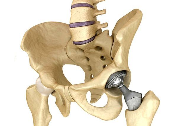
Endoprothetic hip jointsIt is used in severe cases III, when pain syndrome cannot be eliminated, and joint mobility is significantly limited.The prostheses of the hip joint lead to a decrease in pain syndrome, an improvement in the functional status of the joint and the quality of the patient's life.The effect persists for 10-15 years, after which a second operation may be required.During surgery, the hip joint is replaced by the artificial imitation of ceramic, metal (titanium prostheses used more frequently) or polymer.
Forecast.Prevention
The prognosis of the arthrosis of the hip joints in relation to the patient's life is favorable, but the disease often leads to disability.According to the World Health Organization, 80 % of elderly patients with coxarthrosis have a violation of mobility, and 25 % cannot do everyday issues.In this sense, the primary prevention of the osteoarthritis of the hip joints is important.
Preventive measures:
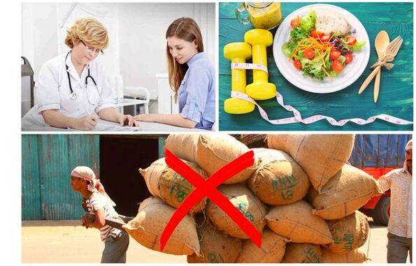
- Reduce body weight.It is necessary to adjust nutrition to reduce the weight and load of the joint.In addition, a decrease in the volume of adipose tissue reduces the amount of inflammation mediators that it released.
- Avoid heavy physical work and sports overloads.Physical overloads are often the cause of the osteoarthritis of the hip joints, while moderate physical activity, on the contrary, improves the condition of the joint cartilage, retains its normal mobility and reduces the load in other joints.
- Correct the underlying disease.If the patient is detected in diseases that can lead to secondary (endocrine, rheumatic and others), the underlying disease is necessary.The normalization of hormonal background and the achievement of persistent remission of rheumatic diseases is the primary prevention of osteoarthritis and allows it to slow down its development.
- Direct a healthy lifestyle.A balanced diet with sufficient content of vegetable and animal protein, polyunsaturated fatty acids and limiting simple carbohydrates, as well as moderate physical activity, avoid the appearance of coxarcherrosis even in the presence of risk factors.
Currently, the prevention of hip articulation diseases is mandatory in neonatology and pediatrics.Over time, the adjusted congenital dysplasia of the hip joint significantly reduces the risk of coxarchesis in adulthood.